

Understanding your biopsy report can feel overwhelming, especially when faced with terms like "atypical" or "suspicious cells." Grasping these concepts not only aids in reducing anxiety but also empowers you to engage meaningfully with your healthcare provider about your next steps. Let's uncover the essential insights that will guide you through this journey.
What You Will Learn
- Atypical Cells: Indicate abnormal appearance but are not definitively cancerous; they could suggest benign conditions or require further investigation.
- Suspicious Cells: Often necessitate additional testing, such as repeat biopsies or imaging studies, due to a higher risk of malignancy.
- Common Causes: Benign conditions like inflammation or hormonal changes can lead to atypical findings, not necessarily indicating cancer.
- Next Steps: Follow-up appointments and further testing are crucial after receiving atypical results to ensure appropriate care.
- Diagnostic Pathways: Understanding the process involving imaging tests and consultations can help alleviate anxiety surrounding results.
- Risk Assessment: Individual factors, such as age and medical history, play a significant role in determining your personal risk of developing malignancy.
- Patient Guidance: Familiarizing yourself with key sections of your pathology report can empower you to have informed discussions with your healthcare provider.
Understanding Atypical Cell Findings in Your Biopsy Report
This visual summarizes the diagnostic pathway and key considerations when atypical or suspicious cells are found in a biopsy report, highlighting common causes, next steps, and specific organ insights.
Atypical Cells vs. Suspicious Cells
- Atypical: Abnormal appearance, not definitively cancer.
- Suspicious: Higher risk of cancer, needs more investigation.
Common Causes
- Benign: Inflammation, infection.
- Other: Aging, hormonal changes.
Diagnostic Pathway: Next Steps
- Follow-Up Appointments: Discuss results & plan.
- Further Testing: Repeat biopsies, imaging (ultrasound, CT, MRI).
- Consultations: Specialist referrals (e.g., oncologists).
- Regular Surveillance: Monitoring over time.
Organ-Specific Insights
- Breast: Atypical ductal hyperplasia, lobular neoplasia.
- Lung: PET scans for nodules.
- Urinary: Cystoscopy, repeat cytology.
Each organ has unique implications requiring specific follow-ups.
Treatment Options
- Monitoring: Careful observation.
- Intervention: Surgery, medications.
- Consultation: Personalized care with specialists.
Understanding Atypical and Suspicious Cells in Your Biopsy Report
When you receive your biopsy report, encountering terms like atypical or suspicious cells can be unsettling. These terms indicate that some cells in your tissue sample do not look like normal cells, but it doesn’t necessarily mean that you have cancer. Understanding what these terms mean can help you navigate your next steps with more confidence.
Atypical cells can present in various forms, and their presence can signify a range of conditions—from benign changes due to inflammation to more serious concerns. It's essential to discuss these findings thoroughly with your healthcare provider, as they can explain the implications specific to your situation.
What Do Atypical Cells Mean in a Biopsy Report?
Atypical cells are those that appear abnormal under the microscope but are not definitively cancerous. These findings might suggest that your healthcare team needs to take a closer look. Suspicious cells, on the other hand, may carry a higher risk of being cancerous, indicating the need for further assessment.
- Atypical Cells: May indicate benign conditions or an early warning sign.
- Suspicious Cells: Often require more extensive investigation, such as repeat biopsies or imaging.
- Communication is Key: Always reach out to your doctor for clarification and next steps.
The key takeaway here is that while these results can feel alarming, they are part of a broader diagnostic process designed to ensure you receive the appropriate care. Remember, every case is unique, so individualized interpretations are crucial!
Common Causes of Atypical Cells in Biopsies
There are several reasons why atypical cells might appear in your biopsy results. Often, benign conditions such as inflammation or infection can lead to atypical findings. For example, chronic inflammation might cause cells to change in appearance, which can confuse the diagnostic process.
- Benign Causes: Conditions like infection or inflammation.
- Aging: As we grow older, our cells can change, which may lead to atypical findings.
- Hormonal Changes: Fluctuations in hormones can also affect cell appearance.
Understanding these common causes of atypical cells helps demystify your biopsy report. It’s important to remember that having atypical cells does not automatically indicate cancer but rather suggests that your healthcare provider needs to conduct further investigations.
Next Steps After Receiving Atypical Results
After you receive a report indicating atypical results, it’s essential to discuss your next steps with your healthcare provider. Recommended follow-up actions often include further testing, like repeat biopsies or specific imaging studies.
- Follow-Up Appointments: Schedule these to discuss your results and next steps.
- Regular Surveillance: Your doctor may recommend monitoring your condition over time.
- Further Testing: Additional biopsies or imaging may be suggested to clarify the diagnosis.
Taking action based on these recommendations can help ensure that you receive the most appropriate care tailored to your specific situation. Always stay engaged and ask questions during these discussions—your understanding is crucial in navigating this process.
Diagnostic Pathways: What to Expect
Once atypical results are confirmed, the diagnostic pathway can become intricate. Your healthcare provider may recommend several next steps, including imaging tests such as ultrasound, CT scans, or MRIs to gather more information about the area of concern. For example, in cases of atypical endometrial hyperplasia, further evaluation is often recommended to rule out more serious conditions.
- Imaging Studies: Used to assess the extent of any abnormalities.
- Additional Biopsies: May be necessary to obtain more tissue for analysis.
- Consultations: Referrals to specialists, such as oncologists, for tailored care.
Understanding the diagnostic process can alleviate some of the anxiety surrounding your results. Keep in mind that your healthcare team is there to support you and guide you through each step of this journey.
Risk Assessment: Understanding Your Individual Probability
Risk assessment is an essential component of understanding what atypical results might mean for you. Your healthcare provider will consider various factors, including your age, medical history, and specific biopsy findings to determine your personal risk of developing malignancy. A comprehensive understanding of your pathology report, as detailed by the American Cancer Society, can provide crucial insights into these risks.
- Statistical Data: Your doctor may share statistics about the likelihood of atypical cells progressing to cancer based on various factors.
- Individual Factors: Age, family history, and previous medical issues play a significant role in risk evaluation.
- Personalized Approach: Each patient’s risk is unique; work with your healthcare provider to understand yours.
Grasping your individual risk can empower you to take proactive steps towards managing your health. Always communicate openly about your concerns and ask for clarification on any uncertainties in your risk assessment.
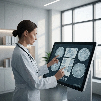
Understanding Histopathology and Cytology in Your Diagnosis
Histopathology and cytology are two essential branches of pathology that help in diagnosing atypical cells. Histopathology involves examining the architecture of tissues under a microscope, while cytology focuses on individual cells. For a deeper dive into these diagnostic processes, resources like those from the National Center for Biotechnology Information offer valuable insights.
- Histopathology: Evaluates tissue samples to determine the presence of disease.
- Cytology: Analyzes cell samples for abnormalities and characteristics.
- Significance of Findings: Both methods provide crucial information to guide diagnosis and treatment.
Understanding these diagnostic tools can help you appreciate the complexity of your report. Collaborate with your healthcare provider to gain insights into how these evaluations fit into your overall diagnosis.

Organ-Specific Insights on Atypical Cells
Atypical Cells in Breast Biopsies
In the context of breast biopsies, atypical cells can have specific implications. Additional tests, such as immunohistochemistry, may be employed to gather further information about the cells in question.
- Common Findings: Atypical ductal hyperplasia or lobular neoplasia are common terms associated with atypical breast results.
- Further Testing: Immunohistochemistry can help in distinguishing between benign and malignant changes.
Understanding these findings can help you have informed discussions with your healthcare provider about your options and next steps.
Atypical Cells in Lung Biopsies
Atypical findings in lung biopsies can be particularly concerning, as they may lead to further investigations like a PET scan to assess any nodules present in the lungs.
- Diagnostics: PET scans can help visualize metabolic activity and detect potential malignancies.
- Nodule Assessment: Follow-up tests are often required to determine the nature of lung nodules.
Being aware of these implications can prepare you for discussions about your lung health and necessary follow-up actions.
Atypical Findings in Urinary Cytology
Atypical cells can also appear in urinary cytology, where they may indicate a need for further evaluation. Follow-up tests might include cystoscopy or repeat cytology to clarify the findings.
- Manifestation: Atypical cells in urine samples can signal urinary tract issues.
- Recommended Follow-Ups: Your healthcare provider might suggest additional tests to ensure comprehensive evaluation.
Understanding potential findings in urinary cytology can help you feel more prepared as you navigate your healthcare journey.
Patient Guidance: Understanding Your Pathology Report
Biopsy reports can be dense and filled with medical jargon. To help you navigate this, I recommend breaking down complex terminology into simpler terms. Key sections to focus on in your pathology report include:
- Cell Type: What type of cells were analyzed?
- Findings: What did the pathologist observe?
- Recommendations: What are the suggested next steps?
Discussing these sections with your healthcare provider can provide clarity and help you understand the implications of your results. Remember, you deserve to have your questions answered!
Exploring Treatment Options for Atypical Cell Findings
Once atypical results are confirmed, exploring treatment options becomes vital. Depending on the findings and potential risks, treatment options can vary widely.
- Monitoring: In some cases, careful observation may be the best approach.
- Intervention: Treatments may include surgical options or medications based on your specific diagnosis.
- Consultation: Engaging with an oncologist can provide personalized care tailored to your needs.
Understanding your options empowers you to make informed decisions about your health care. Always engage with your healthcare team to determine the best course for your unique situation.
FAQs: Common Concerns About Atypical Cells
Receiving a biopsy report with atypical findings raises many questions. Here are some common concerns and answers:
- What do atypical cells mean for my health? Atypical cells may require further assessment, but they do not definitively indicate cancer. They suggest that cells appear abnormal and need closer monitoring or investigation to determine their nature.
- How often should I follow up after an atypical result? Follow-up intervals depend entirely on your specific case, the type of atypicality found, and your personal risk factors. Your healthcare provider will give you tailored advice on the appropriate monitoring schedule.
- Will I definitely need more tests if atypical cells are found? In most cases, yes. Atypical findings often lead to recommendations for additional testing, such as repeat biopsies, specialized imaging (e.g., MRI, CT scans, PET scans), or referral to a specialist, to clarify the diagnosis and guide further management.
- Can atypical cells turn into cancer? Some types of atypical cells, especially those classified as "suspicious," do have a higher potential to progress to cancer over time. However, many atypical findings remain benign or resolve on their own. The risk of progression depends on the specific type of atypicality and other individual factors.
- What are the common causes of atypical cells besides cancer? Atypical cells can result from various benign conditions, including inflammation, infection, hormonal changes, and the natural aging process of cells. These non-cancerous causes often lead to temporary cellular changes that mimic more serious conditions.
Reaching out to your healthcare provider with these questions can foster a proactive approach to your health. Stay informed and engaged as you navigate this process!
We Want to Hear From You!
What is your biggest concern when it comes to understanding your biopsy results? Share your thoughts below:
Final Thoughts on Atypical and Suspicious Cells in Biopsy Reports
Receiving a biopsy report with atypical or suspicious cells can be daunting. However, understanding what these terms mean is the first step toward taking control of your health. Remember, atypical results do not automatically indicate cancer; they require a thorough and individualized assessment. It's crucial to engage proactively with your healthcare providers to clarify the implications of your results and discuss next steps.
As you navigate this process, I encourage you to ask questions and express any concerns. Open communication can lead to a better understanding of your situation and the options available to you. Empower yourself with knowledge and seek clarity on your biopsy findings, as this can significantly alleviate anxiety and foster confidence in your healthcare journey.
Resources for Further Support and Information
To help you further in your journey, I’ve gathered some valuable resources that you can utilize. These materials are designed to support you through understanding your biopsy results and the next steps you may need to take. Here are some key resources:
- Downloadable Patient Checklists: A handy checklist to keep track of your questions and necessary follow-up actions.
- Informational Guides: Detailed articles on biopsy types, what to expect, and explanations of medical terminology.
- Support Groups: Connect with others who are going through similar experiences and can offer support.
- Specialist Referrals: Access to clinics and oncologists who specialize in interpreting biopsy results and providing personalized care.
At What Is A Biopsy, we are committed to ensuring that you have the information you need to navigate your healthcare decisions confidently. Don’t hesitate to reach out for further clarification or support—your health is worth it!
Recap of Key Points
Here is a quick recap of the important points discussed in the article:
- Atypical Cells: Indicate abnormal appearances that may suggest benign conditions or early signs of concern.
- Suspicious Cells: Often require further investigation, such as repeat biopsies or imaging, to clarify results.
- Follow-Up Actions: Regular appointments and possibly additional testing are crucial after receiving atypical results.
- Understanding Risks: Individual risk assessments based on age, medical history, and biopsy findings are essential.
- Communication with Healthcare Providers: Always ask questions and clarify terms on your biopsy report to better understand your health.